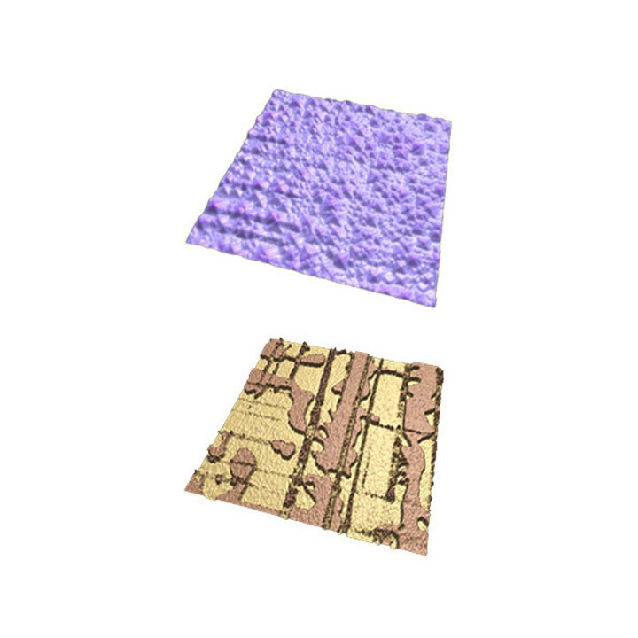
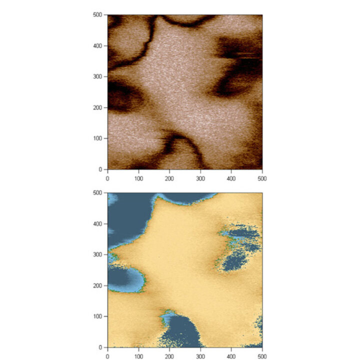
Tooth Dentin
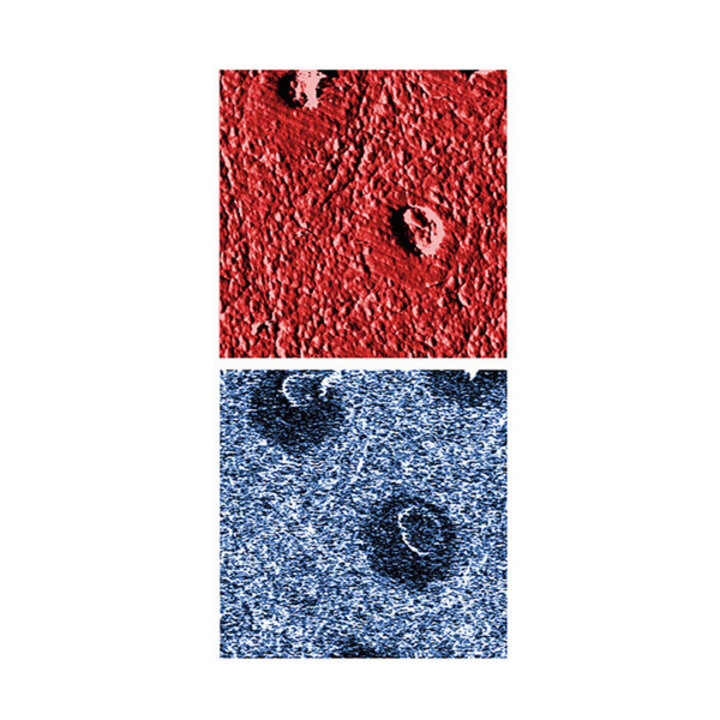
Topographic (top) and PFM amplitude (bottom) of tooth dentin.
Scan size is 10 x10 µm2.
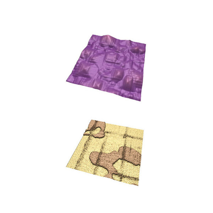
Collagen Fibers
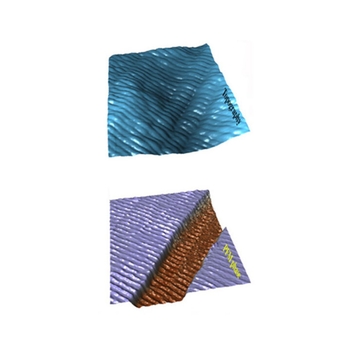
Topographic (top) and PFM phase (bottom) of collagen fiber.
Scan size is 2 x 2 µm2.
Sample courtesy of G. Fantner.
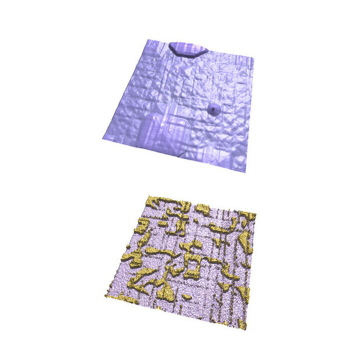
Domain Kinetics
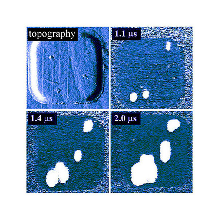
PFM images of instantaneous domain configurations developing during
polarization reversal in epitaxial PZT capacitors. Scan size is 2.5 x 2.5 µm.
Sample courtesy of M. Alexe (MPI, Halle).
Butterfly Wing
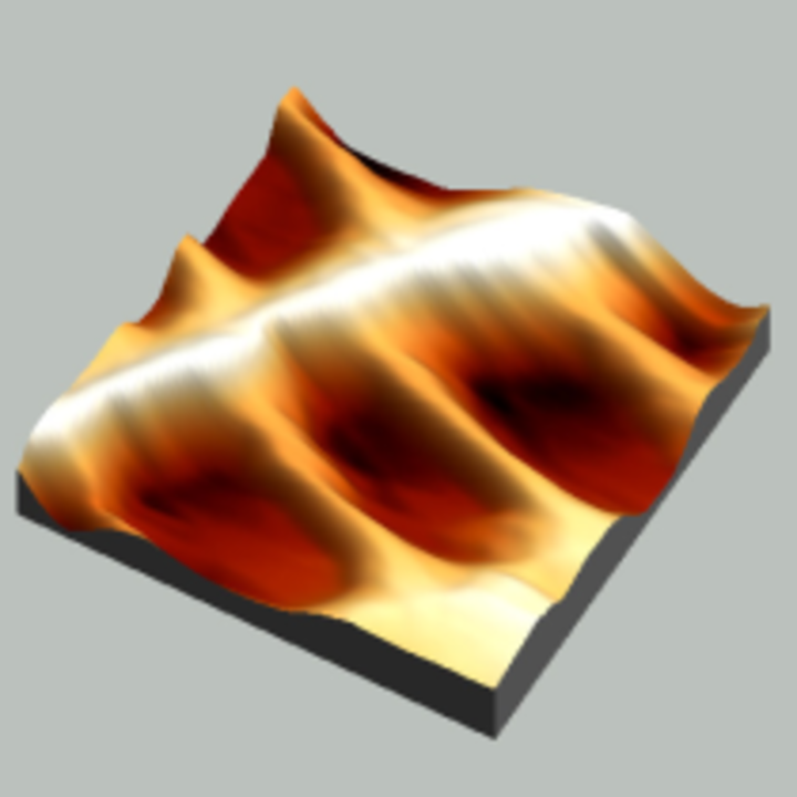
Perspective view of butterfly Vanessa Virginiensis wing scale.
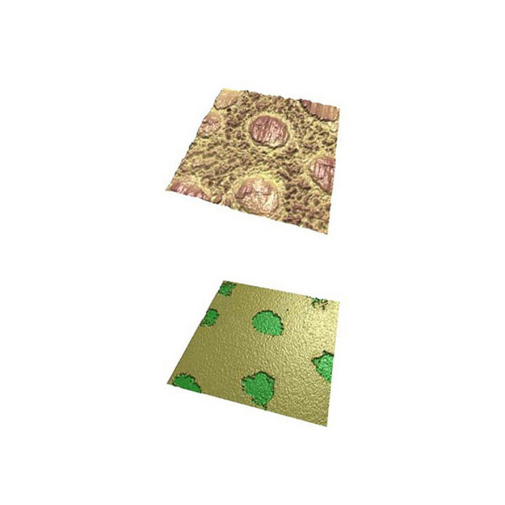
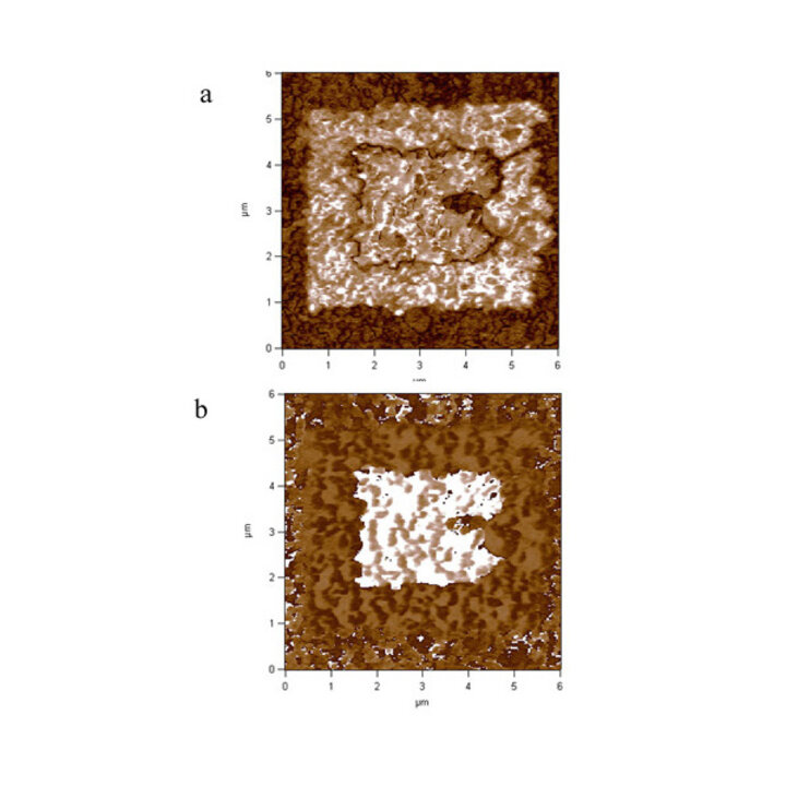
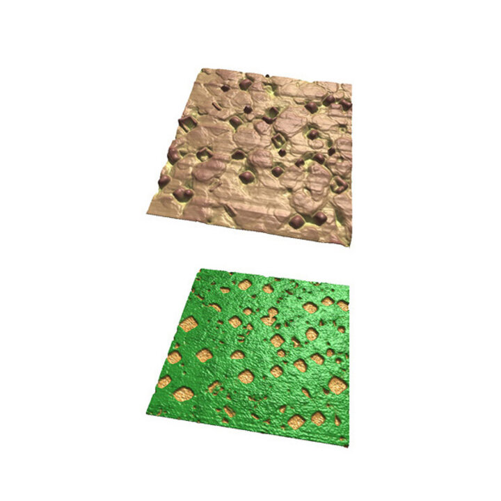
FeRAM
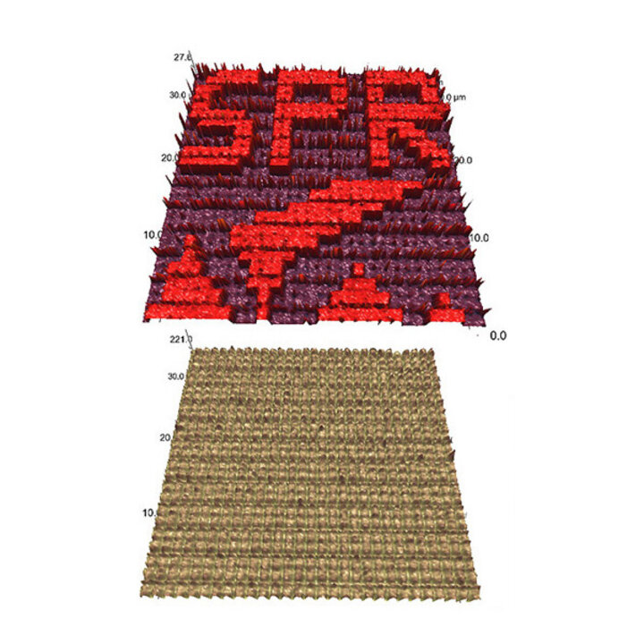
PFM (top) and topograpic (bottom) images of microscale capacitor array.
Sample courtesy of Toshiba.
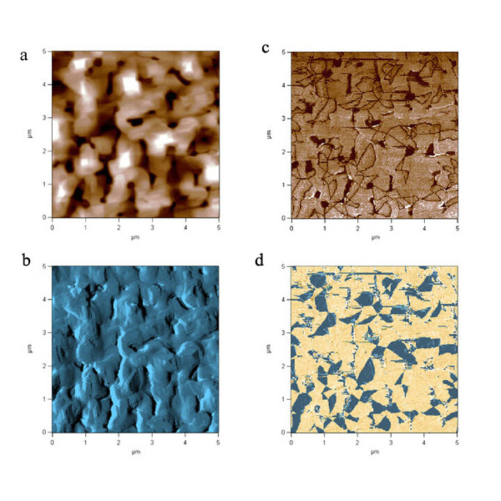
Lead Titanate pt 1
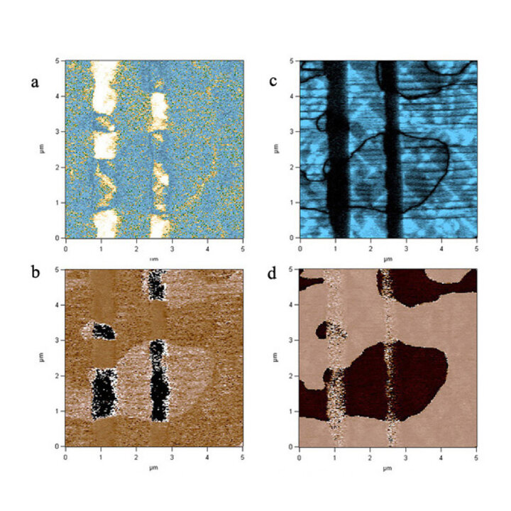
Lateral PFM (a-amplitude, b-phase) and vertical PFM (c-amplitude, d-phase)
images of a-c-domain structure in PbTiO3 film.
Sample courtesy of H. Funakubo (TIT).
Lead Titanate pt 2
Lateral PFM (a-amplitude, b-phase) and vertical PFM (c-amplitude, d-phase)
images of a-c-domain structure in PbTiO3 film.
Sample courtesy of H. Funakubo (TIT).
Lead Titanate pt 3
Topographic and PFM images of a-c-domain structure in PbTiO3 film.
Scan size is 1.5 x 1.5 µm2.
Sample courtesy of H. Funakubo (TIT).
Lead Titanate pt 4
Topography (top) and PFM phase (bottom) images of a-c-domain structure
in PbTiO3 film. Scan size 20 x 20 µm2.
Sample courtesy of H. Funakubo (TIT).
Lead Titanate pt 5
Topography (top) and PFM phase (bottom) images of a-c-domain structure
in PbTiO3 film. Scan size 5 x 5 µm2.
Sample courtesy of H. Funakubo (TIT).
Lead Titanate pt 6
Topography (top) and PFM phase (bottom) images of a-c-domain structure
in PbTiO3 film. Scan size 20 x 20 µm2.
Sample courtesy of H. Funakubo (TIT).
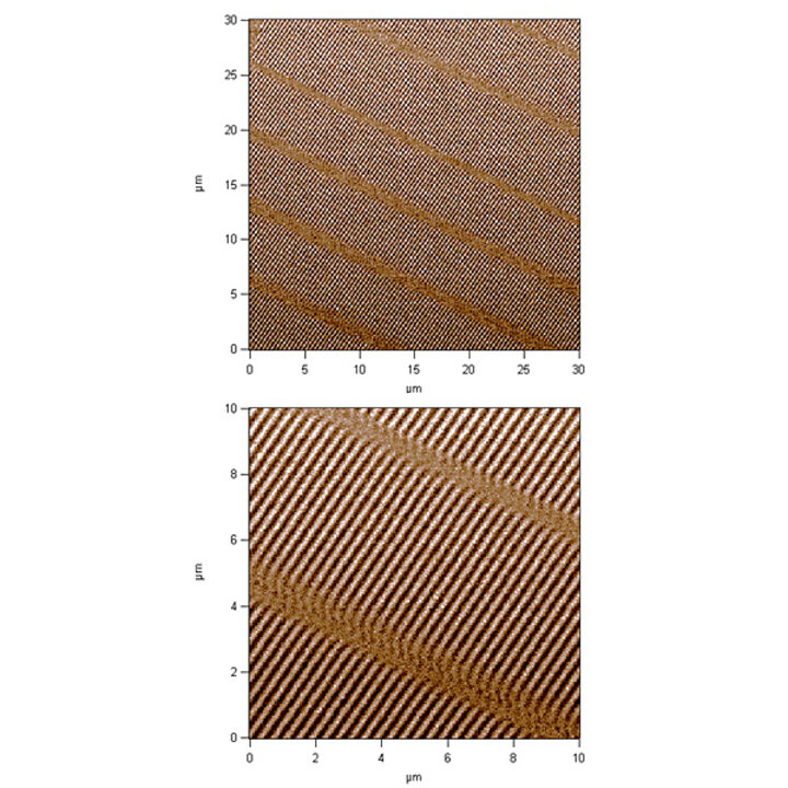
Lead Titanate pt 7
Topography (top) and PFM phase (bottom) images of c-domain structure
in PbTiO3 film. Scan size 20 x 20 µm2.
Sample courtesy of H. Funakubo (TIT).
Domain Writing
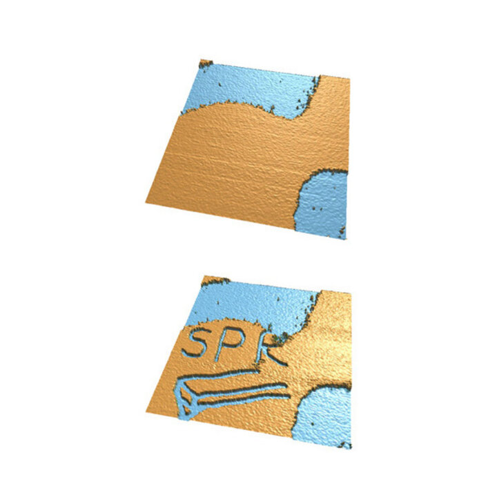
PFM phase images of initial (top) and modified (bottom) domain structure
in PbTiO3 film. Scan size 10 x 10 µm2.
Sample courtesy of H. Funakubo (TIT).
PVDF pt 1
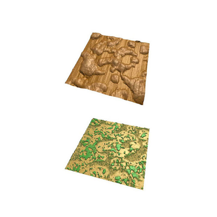
Topographic (top) and PFM phase (bottom) images of PVDF film.
Scan size is 1 x 1 µm2.
Sample courtesy of T. Reece and S. Ducharme (UNL).
PVDF pt 2
PFM Amplitude (top) and phase (bottom) images of PVDF film.
Scan size is 500 x 500 nm2.
Sample courtesy of T. Reece and S. Ducharme (UNL).
Poled PVDV
PFM amplitude (top) and phase (bottom) images of poled PVDF film.
Sample courtesy of T. Reece and S. Ducharme (UNL).
BFCO
Topographic (top) and PFM phase (bottom) images of domain structure
in BFCO thin film. Scan size is 5 x 5 µm2.
Sample courtesy of H. Funakubo (TIT).
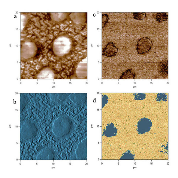
Bismuth Ferrite
a-Topographic, b-deflection, c-PFM amplitude and d-PFM phase images of BFO thin film.
Sample courtesy H. Funakubo (TIT).
Hard Disk
MFM images of a hard disk.